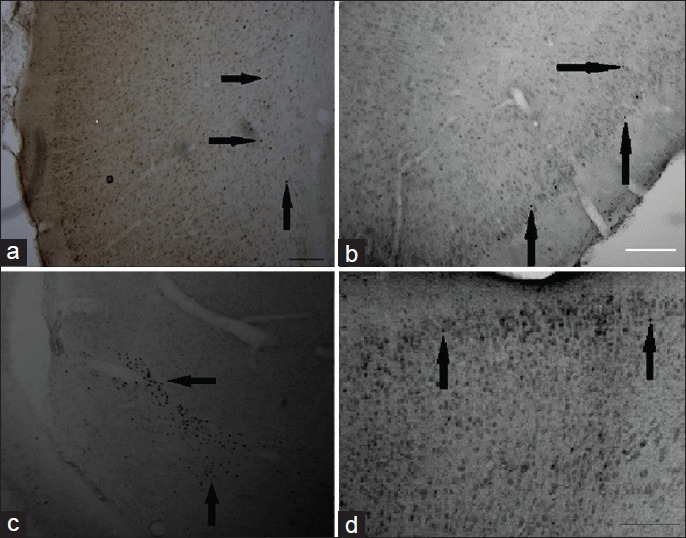
Figure 1.

Representative photomicrographs of cFos and BDNF expression in the cortex and hippocampus at 1 and 3 days EW. Immunohistochemistry using cFos antibody showed an effect of EW on expression of cFos labeled with DAB. Generally, EW rats showed a greater expression of cFos compared to pair-fed controls. No staining was observed when either the anti-Fos antibody or the secondary antibody was omitted from the experiment. (a) Control female at 24 hour EW, piriform cortex; (b) EW female at 24 hours, piriform cortex; (c) EW OVX at 3 day, hippocampus, using cFos antibody; (d) EW female at 3 day EW, motor cortex using BDNF immuno-labeling; Magnification: ×40. Scale bars: A-C, 200 mm; D, 100 mm. Magnification: ×40. Scale bars: A, B: 200 mm
