Figure 2.
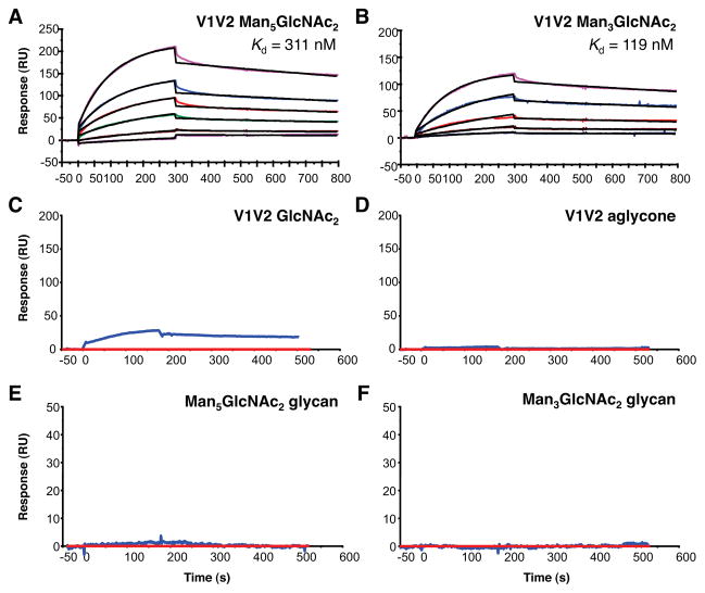
Binding of mAb PG9 to gp120 V1V2 glycopeptides. SPR sensorgrams showing binding of mAb PG9 to V1V2 glycopeptides derivatized with Man5GlcNAc2 (A) and Man3GlcNAc2 (B). V1V2 Man5GlcNAc2 binding curves are shown for glycopeptide concentrations at 5, 10, 20, 30 and 40 μg/mL and V1V2 Man3GlcNAc2 at 1, 2, 5, 10 and 20 μg/mL. Control SPR sensograms showing minimal to no binding of mAb PG9 to V1V2 GlcNAc2 (C), V1V2 aglycone (D), Man5GlcNAc2 glycan alone (E), and Man3GlcNAc2 glycan alone (F). V1V2 GlcNAc2 and aglycone peptides were injected at 200 μg/mL (C, D) and Man5GlcNAc2 and Man3GlcNAc2 glycans at 25 μg/mL (E, F) over PG9 captured on anti-human IgG (Fc-specific) surfaces. SPR data were derived following subtraction of non-specific signal on a control anti-RSV mAb (Synagis, red curve in C–F).

