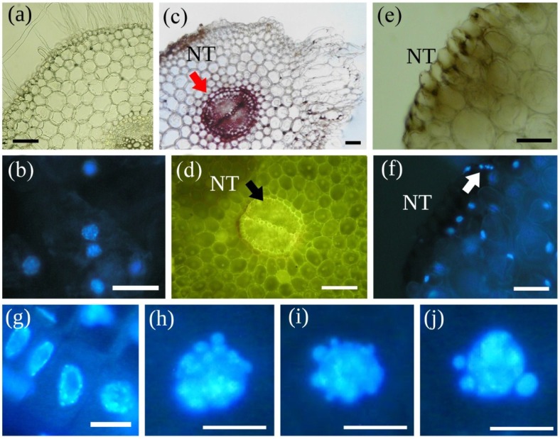
Figure 2.
MCY-LR induces callus formation (swollen cells), PCD and necrosis in the rhizodermis and cortex of S. alba primary roots. (a) Control root collar; (b) Same tissues stained with DAPI; (c) Cross-Section of main root-hypocotyl transition zone of seedlings treated with 20 μg mL−1 MCY-LR, showing the formation of a callus-like tissue (CA), necrosis (NT) and intensive lignification of endodermis and stele as shown by phloroglucin-HCl staining (arrow); (d) High autofluorescence of inner root tissues (arrow) induced by 20 μg mL−1 MCY-LR exposure: autofluorescence fades away in necrotic tissue (NT); (e) 20 μg mL−1 MCY-LR induces necrosis of rhizodermis and adjacent tissues (NT); (f) Same tissues as in (e) stained with DAPI, nuclei are absent in necrotic cells (NT), and the fragmentation of nuclei can be observed in cells neighboring necrotic tissue (arrow); (g–j) Nuclei from the root tip meristem-elongation zone transition; (g) control; (h–j) Treatment with 5 μg mL−1 MCY-LR: nuclear blebbing (h,i) leading to the fragmentation of chromatin (j); Scalebars: 80 μm (a,b,e,f), 200 μm (c,d) and 15 μm (g–j). Micrographs taken by M. M-Hamvas (a,c,e), C. Máthé (b,d,f–j).

