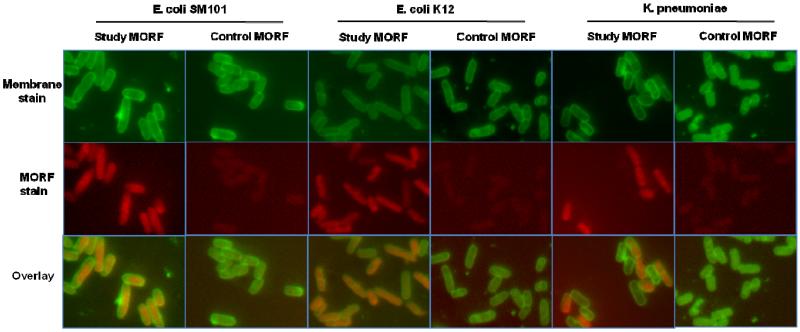
Fig. 5.
Fluorescence microscopy images showing the accumulation of study and control MORFs in live E. coli (SM101 and K12) and K. pneumoniae after 2 h. Top row shows the cell membrane stained with FM1-43, middle row shows MORFs conjugated with AF633, and bottom row shows overlay of the two colors. (Magnification 1000 ×).

