Figure 2.
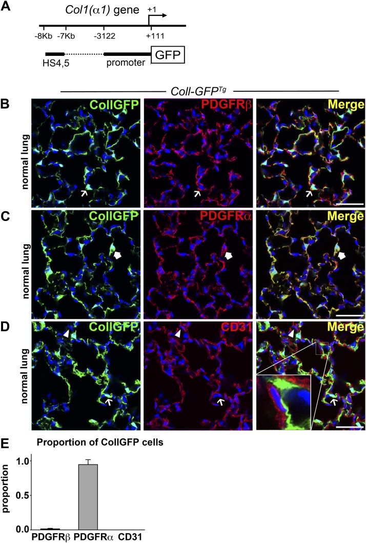
Collagen-Iα1 GFP transgene–expressing cells coexpress PDGFRα in normal lung. (A) Schema showing the Coll-GFP transgene with a 3.2-kb fragment of the Col1a1 promoter and a 1-kb enhancer fused to GFP. (B–D) Confocal images showing the expression of GFP in the adult lung of Coll-GFPTg mice and colabeling with (B) PDGFRβ, (C) PDGFRα, and (D) CD31. Example of Coll-GFP+ cell (green) lacking the marker is indicated by a thin arrow. GFP− cell expressing the indicated marker (red) is indicated by an arrowhead. Coll-GFP+ cell coexpressing the indicated marker (yellow plasma membrane in merged image) is indicated by a thick arrow. Inset in D shows a space separating Coll-GFP+ cell from the endothelium. (E) Graph showing the proportion of Coll-GFP cells coexpressing the indicated markers. Bar = 50 μm. Mean ± SEM. n = 3 mice per group.

