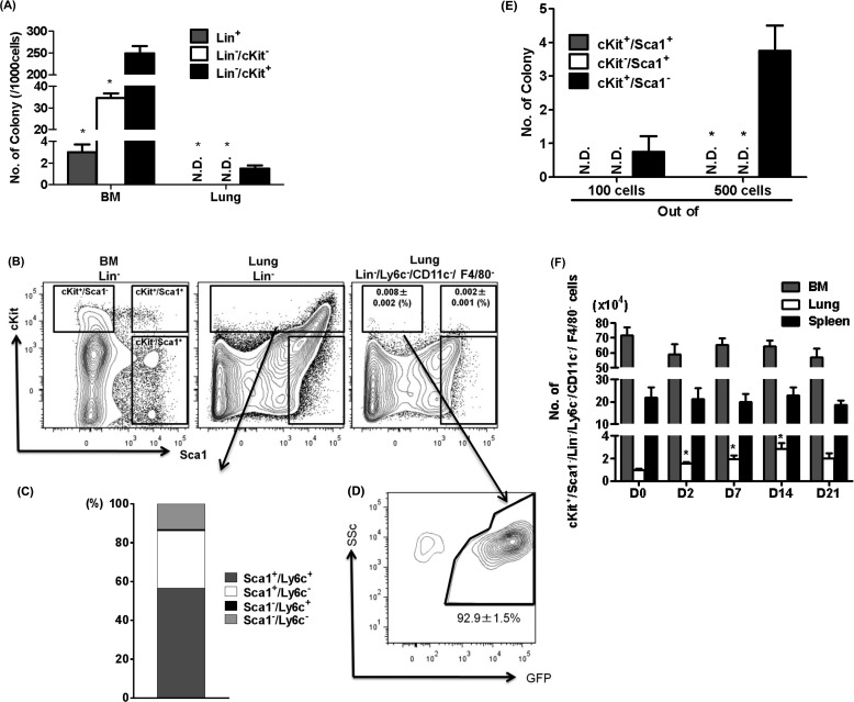
Figure 4.
Characteristics of colony-forming cells in the lung. (A) Using Methocult colony forming assay, numbers of colony derived from Lin+, Lin−/cKit−, or Lin−/cKit+ cells in the bone marrow (BM) and lung were counted (n = 4 per group). *P < 0.05 versus Lin−/cKit+. (B) Flow-cytometric analyses of Sca1 and cKit expression in BM Lin− cells, lung Lin− cells, or lung Lin−/Ly6c−/CD11c−/F4/80− cells are shown. The numbers of cKit+/Sca1−/Lin−/Ly6c−/CD11c−/F4/80− and cKit+/Sca1+/Lin−/Ly6c−/CD11c−/F4/80− cells in lung were enumerated and shown as percent of total lung cells (n = 4 per group). (C) The numbers of Sca1 and Ly6c positivity in lung Lin−/cKit+ cells were enumerated and shown as percent of total. (D) Numbers of colonies derived from sorted cKit+/Sca1+, cKit−/Sca1+, or cKit+/Sca1− cells among the lung Lin−/Ly6c−/CD11c−/F4/80− cells are shown. *P < 0.05 versus cKit+/Sca1−. (E) GFP positivity rate in lung cKit+/Sca1−/Lin−/Ly6c−/CD11c−/F4/80− cells obtained from BM-chimera mice are shown (n = 4 per group). (F) Kinetics of cKit+/Sca1−/Lin−/Ly6c−/CD11c−/F4/80− cells in the BM, lung, and spleen revealed significant increases occurred only in the lung in the BLM-treated mice (n = 3–5 per group). *P < 0.05 versus D0.

