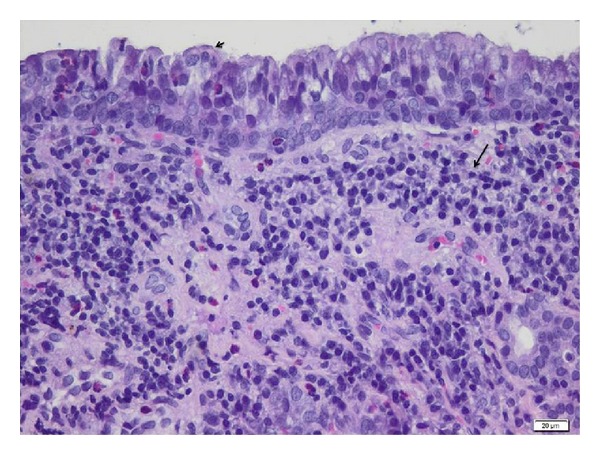
Figure 2.

Mucosa around the ostia at the moment of surgical intervention-light microscopy. Maxillary sinus biopsy showing thickening of sinus mucosa, lymphoid follicle hyperplasia, cilia degeneration (black arrow head), diffuse and several infiltrate with mononuclear cells (black arrow), heterophils, and scattered eosinophils. H&E stain, bar = 20 μm.
