Figure 3.
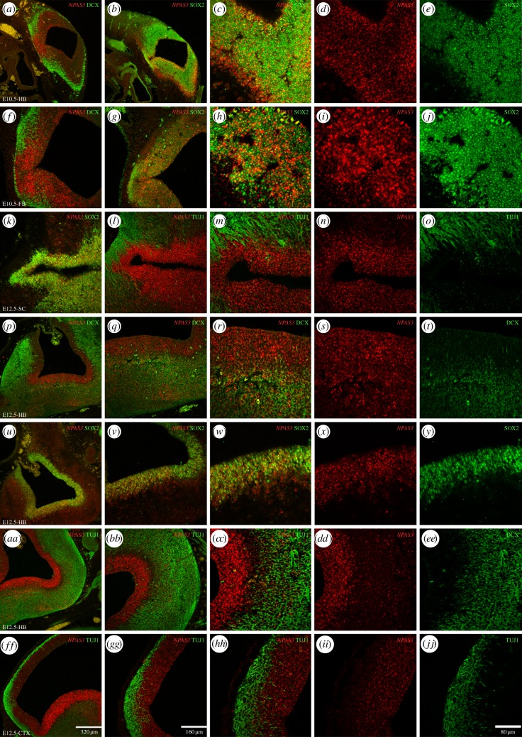
NPAS3 expression in the developing mouse brain. The expression of NPAS3 is shown through ISH in combination with immunohistochemical detection of the early neuronal markers DCX and β III-tubulin (TUJ1) and the undifferentiated NPC marker SOX2 in cryostat sections of E10.5 and E12.5 mouse embryos. Expression of NPAS3 at E10.5 is shown in the hindbrain (a–e) and the forebrain (f–j) in combination with DCX (a,f) and SOX2 (b–e and g–j). At E12.5 expression is shown in the spinal cord (k–o), the hindbrain (p–y and aa–ee) and the forebrain (ff–jj). In the spinal cord, NPAS3 is shown in combination with SOX2 (k) and TUJ1 at low (l) and high magnification (m–o). In the hindbrain, expression is shown in combination with DCX (p–t), SOX2 (u–y) and TUJ1 (aa–ee). In the forebrain, the expression is shown in combination with TUJ1 at low (ff,gg) and high magnification (hh–jj).

