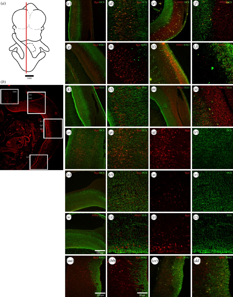
Figure 4.
β-Galactosidase expression pattern at E12.5 in different cellular types in the developing brain. (a) Schematic of an E12.5 whole embryo where a red line indicates the approximate location of the histological section located below. (b) Low-resolution image of a histological section of a transgenic mice expressing β-galactosidase under the control of the 2XHAR142-Pt sequence in the hindbrain and spinal cord. White boxes over the image indicate approximate location of the high-resolution images shown on the right. High-resolution confocal photographs at the right show the expression pattern of the reporter gene lacZ protein product β-galactosidase (bgal) at different levels of the spinal cord (c,d,g,h,k,l,o,p) and the hindbrain (s–z and aa–bb). The expression of β-galactosidase is shown in combination with the expression of the early neuronal markers DCX and β III-tubulin (TUJ1) and the undifferentiated NPC marker SOX2 in the developing central nervous system. The expression of NPAS3 in combination with the same cellular type markers is shown for some cases at approximate similar levels in the spinal cord (e,f,i,j,m,n) and the hindbrain (cc,dd).

