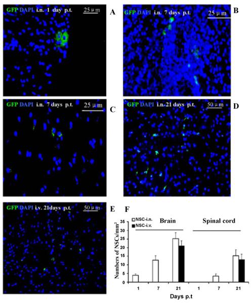
Figure 2. Localization and migration of transplanted GFP-aNSCs in the CNS.
Mice treated with aNSCs i.n. and i.v. were sacrificed at days 1, 7 and 21 p.t., and brains and spinal cords were harvested for immunohistology. At day 21 p.t., all groups were examined in the same region of the corpus callosum and spinal cord. Transplanted aNSCs (green) from i.n. delivery were primarily confined to brain perimengingeal sites at day 1 p.t. (A), but not spinal cord (data not shown), while at day 7 p.t. these cells were found in both brain (B) and spinal cord (C). At day 21 p.t., the majority of these cells from both i.n. (D) and i.v. (E) had migrated to the parenchyma of the corpus callosum (not shown) and spinal cord. Nuclei are stained with DAPI (blue). (F) Quantitative analysis of GFP-aNSCs that reached the parenchyma of EAE at days 1, 7 and 21 p.t. Symbols represent mean values and SD of 6–8 mice from each group. *p<0.05, comparison between 7 and 21 days p.t.; #p<0.05, comparison between i.n. delivery and i.v. injection.

