Figure 2.
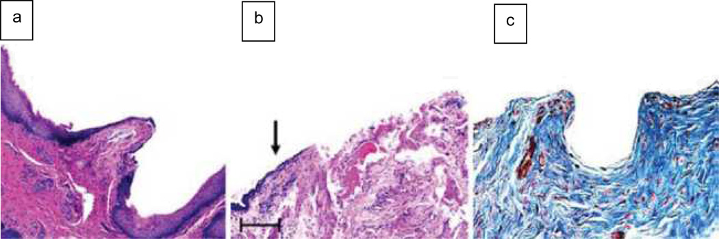
Histology comparing short and long urethral grafts. (a) Hematoxylin and eosin staining of the anastomosis between normal urethral tissue (NU) and the acellular graft (GR) in the 0.5 cm defect, which indicates ingrowth of normal cell types. (b) Representative cross section of a longer graft. The arrow points to increased fibrosis and a friable epithelial surface, beginning approximately 0.5 cm from the tissue-to-graft anastomosis. (c) Masson’s trichrome staining of a longer graft confirms the increased collagen deposition that is characteristic of fibrosis.

