Figure 1.

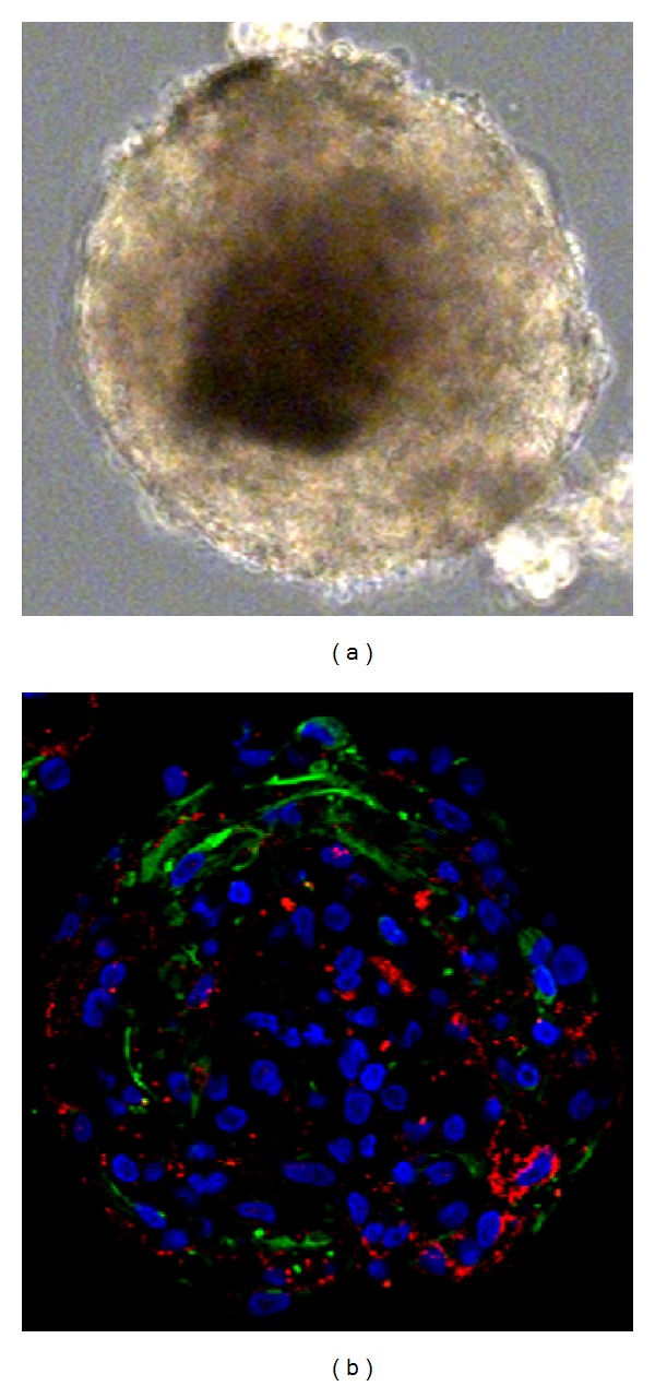
Light microscopy of secondary sphere derived from postmortem CE (a). Immunocytochemical analysis of secondary spheres derived from postmortem CE (b), costaining of Nestin (green) and Claudin-1 (red). Nuclear staining with Hoechst (blue).

Light microscopy of secondary sphere derived from postmortem CE (a). Immunocytochemical analysis of secondary spheres derived from postmortem CE (b), costaining of Nestin (green) and Claudin-1 (red). Nuclear staining with Hoechst (blue).