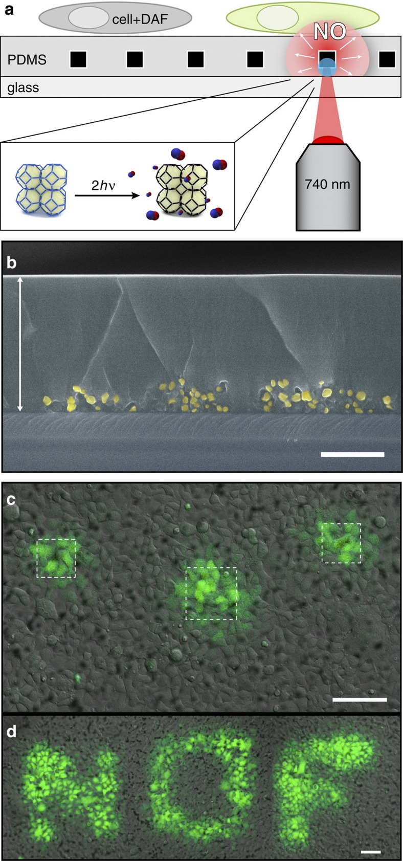
Figure 4. Spatiotemporally controlled release of NO.
(a) Schematic illustration of the localized cell-stimulation platform. The PDMS-embedded NOF-1 crystals are locally photoactivated by two-photon near-infrared laser irradiation. The generated NO diffuses through the PDMS layer and reacts with an intracellular NO fluorescent indicator, DAF-FM. (b) Cross-section scanning electron microscopy image of NOF-1/PDMS substrate. The white arrow highlights the homogeneous PDMS layer. NOF-1 crystals (yellow) are localized on the bottom part of the substrate, ensuring their isolation from the cellular medium (scale bar, 10 μm). (c) Confocal microscopy images of NOF-1-embedded substrates cultured with HEK293 cells introduced via DAF-FM. The selective photoactivation of the NOF-1 crystals (white squares) induced a fluorescent response in the surrounding cells, highlighting the localized NO delivery and uptake (scale bar, 100 μm). (d) Further demonstration of spatiotemporal control by writing ‘NOF’ upon activation of the selected regions (scale bar, 100 μm).

