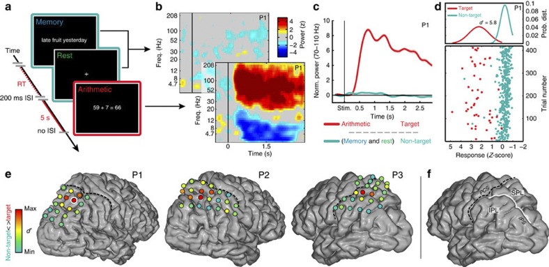
Figure 1. ECoG responses in the human IPS region during experimental arithmetic condition.
The experimental task (a) consisted of self-paced arithmetic (simple addition equations) and non-arithmetic conditions (memory statements and 5-s fixation). During the arithmetic condition, we found a significant increase of activity in some but not all electrodes. As seen in the time-frequency plot (b), this activity was best measured in the high-frequency broadband (HFB, 70–110 Hz) range. The temporal profile of relative HFB power changes (c) are shown for one representative electrode (P1–1) averaged across trials. Using d′-values, the separation of the distributions of HFB responses during arithmetic (red, target) and non-arithmetic (blue, non-target) conditions (d) could be quantified. In each participant’s brain, the d′-values were mapped (e), and the sites with the highest d′-values (red) were located around the IPS region (white line in f). The IPS divides the superior (SPL) from the inferior parietal (IPL) lobules. PCS (post central sulcus) is shown with dashed line. Among all subjects, electrode P1–1 (red electrode with white circle in e) had the highest d′-value.

