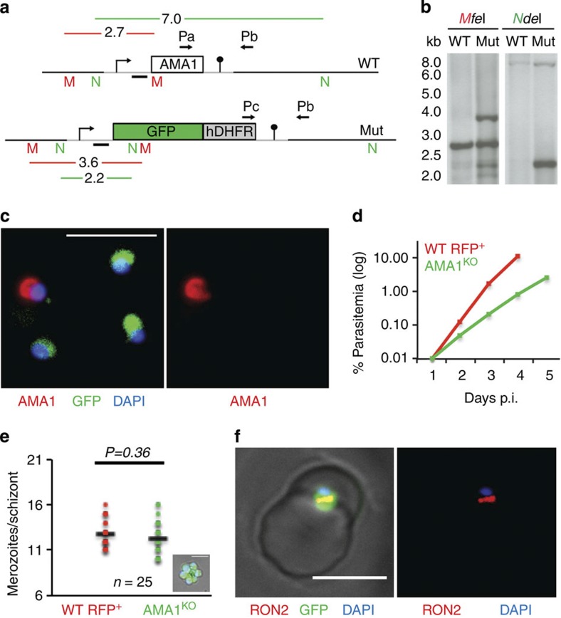
Figure 4. Selection in vivo of P. berghei AMA1KO merozoites.
(a) Schematic illustration of the knockout strategy. Grey lines represent regions of homology for recombination. Integration of the targeting sequence disrupts the AMA1-coding sequence and replaces it by fluorescent (GFP) and drug resistance (hDHFR) markers. Pa, Pb and Pc: primers used for diagnostic PCR in Fig. 8d. M, MfeI and N, NdeI: enzymes used for digestion before Southern blotting. Southern blotting probe is represented as a black line. (b) Southern blot analysis of the AMA1 locus in wild-type (WT) ANKA strain and mutant (Mut) parasites. Digestion with the enzymes MfeI or NdeI produces fragments of different sizes for WT or mutant loci. (c) AMA1 immunostaining of GFP+ (AMA1KO) and GFP− (pyrimethamine-resistant WT) merozoites. Scale bar, 10 μm. (d) Kinetics of in vivo growth of GFP+ (AMA1KO) and WT RFP+ (WT RFP) in co-infected mice. Parasitemia was assessed daily by FACS and plotted as the mean parasitemia measured in five mice. Data are representative of two independent experiments. (e) Number of merozoites per schizont formed 16 h after in vitro incubation of erythrocytes infected with GFP+ (AMA1KO) or WT RFP+ ring stages. Only cells infected with a single parasite were counted. A picture of a cell infected with a GFP+ (AMA1KO) schizont is shown in the plot. Scale bar, 5 μm. P=0.36 in one-way analysis of variance followed by Tukey HSD test. (f) RON2 immunostaining of a GFP+ (AMA1KO) merozoite invading a host cell shows the formation of a RON2 ring, suggestive of a formed TJ. Scale bar, 5 μm. DAPI, 4′,6-diamidino-2-phenylindole.

