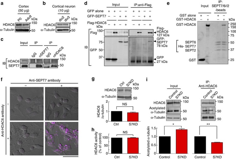
Figure 5. Physiological and direct interaction between septins and the major tubulin deacetylase HDAC6.
(a,b) Immunoblot for endogenous HDAC6 (a) in newborn (P0) and adult cerebral cortex and (b) in primary cerebrocortical neurons at div2 and 3. Each lane respectively contained 50/10 μg total protein. (c) Mutual co-immunoprecipitation of endogenous SEPT7 and HDAC6. Lysates from div2 cerebrocortical neurons were incubated with protein A beads coated with nonimmune IgG, anti-SEPT7 antibody or anti-HDAC6 antibody, and these proteins were detected by immunoblot in a reciprocal manner. (d) Co-expression and mutual co-immunoprecipitation of GFP-SEPT7 and Flag-HDAC6 in heterologous cells. (Left) Anti-Flag antibody detected Flag-HDAC6 (and gave nonspecific faint bands in the 1st and 3rd lanes) and anti-GFP antibody detected GFP and GFP-SEPT7. (Right) When Flag- HDAC6 was captured on anti-Flag beads, GFP-SEPT7 was co-immunoprecipitated, but GFP was not (compare the 2nd and 4th lanes. *, IgG light chain). (e) Direct binding between the purified, recombinant septin complex and HDAC6 in vitro. Immobilized His-tagged SEPT7/6/2 captured GST-HDAC6 but not GST alone. (Coomassie blue staining). (f) Representative results of in situ proximity ligation assay for endogenous SEPT7 and HDAC6 in div2 cerebrocortical neurons. Fluorescent puncta were generated in the somata and neurites only when anti-SEPT7 antibodies and anti-HDAC6 antibodies were present in close proximity. Scale bars, 25 μm. (g) Immunoblot showing that SEPT7 depletion did not affect the amount of HDAC6 in div2 cerebrocortical neurons. (Triplicated experiment. NS, P>0.05 by t-test). Error bars denote s.e.m. (h) SEPT7 depletion via RNAi (#1, S7KD) did not alter the deacetylating activity of HDAC6. Cell lysates as in (b) were subjected to an in vitro assay with a fluorogenic substrate. (Triplicated experiment. NS, P>0.05 by t-test). Error bars denote s.e.m. (i) SEPT7 depletion via RNAi (#1, S7KD) caused dissociation of HDAC6 and acetylated α-tubulin. (Left) Cell lysates as in (b,h) were immunoblotted for endogenous HDAC6, acetylated α-tubulin and total α-tubulin. Hyperacetylation of α-tubulin in SEPT7-depleted neurons was recapitulated (cf. Fig. 3d). Each lane contained 50 μg total protein. (Right) Although SEPT7 depletion increased acetylated α-tubulin, its association with HDAC6 was paradoxically reduced. (Triplicated experiments. *P<0.05, **P<0.01 by t-test). Error bars denote s.e.m.

