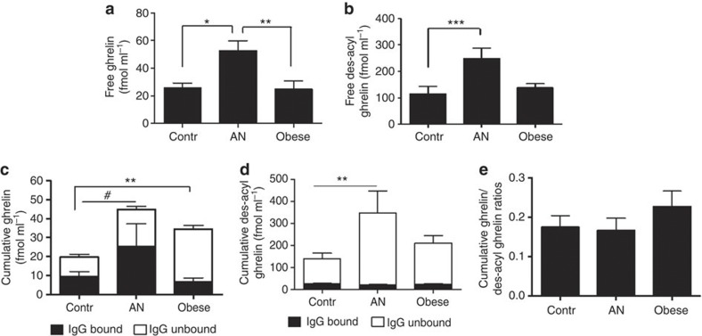
Figure 1. Ghrelin and des-acyl ghrelin concentrations in humans.
Plasma concentrations of ghrelin (a) and des-acyl ghrelin (b) before IgG extraction. Ghrelin (c) and des-acyl ghrelin (d) bound to the plasma-extracted IgG or IgG unbound assayed in IgG-deprived effluents of the same plasma samples are shown in the same bar as cumulative ghrelin concentrations for which statistical analysis is presented. Ratios of cumulative plasma concentrations between ghrelin and des-acyl ghrelin (e). (a) Kruskal–Wallis (K–W) test, P=0,002, Dunn’s *P<0.05, **P<0.01; (b) K–W test, P=0.0008, Dunn’s ***P<0.001; (c) K–W test, P=0.001, Dunn’s **P<0.01, Mann–Whitney test #P<0.05; (d) K–W (M-W) test, P=0,009, Dunn’s **P<0.01. (Contr. n=14, AN n=12 and obese n=14, error bars, s.e.m.).

