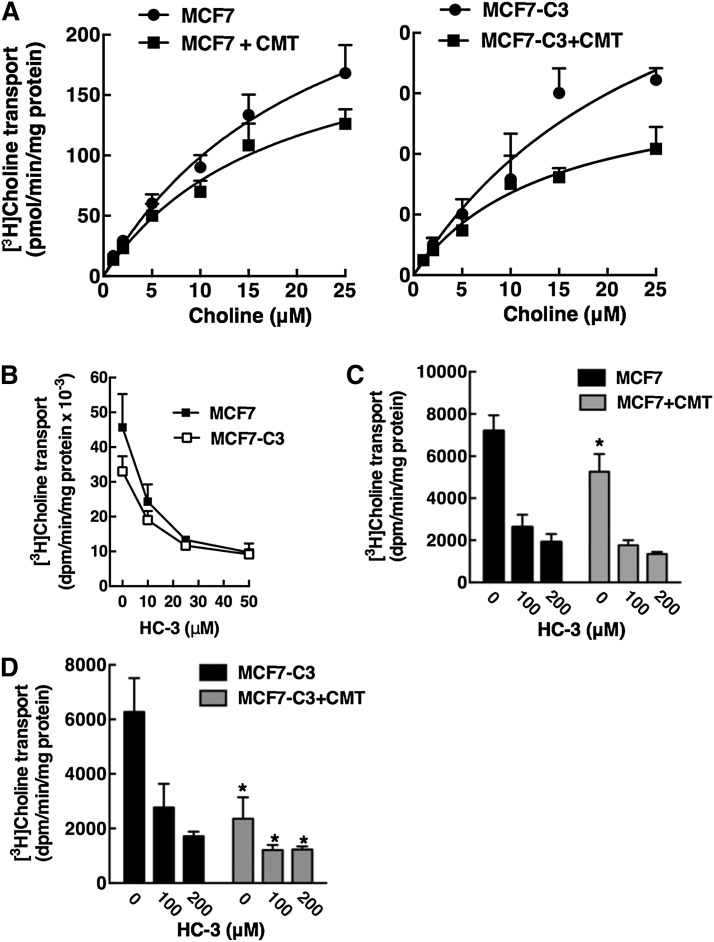
Fig. 10.
Choline transport activity is reduced in apoptotic MCF7 and MCF7-C3 cells. A: MCF7 and MCF7-C3 cells were treated with camptothecin (CMT, 15 μM) for 24 h. Cells were subsequently rinsed with Krebs-Ringer buffer and uptake of increasing concentrations of [3H]choline (1–25 μM) was measured at 37°C for 10 min as described in Materials and Methods. KD and Bmax values were determined by Scatchard analysis fit to a single binding site model. Results are the mean and standard deviation of triplicate measurements from five experiments. B: Uptake of 10 nM [3H]choline into MCF7 and MCF7-C3 cells was assayed for 10 min at 37°C in the presence of increasing concentrations HC-3. Results are the mean and standard deviation of three experiments. C, D: Uptake of 20 μM [3H]choline into MCF7 and MCF7-C3 cells (treated with control solvent or 15 μM CMT for 24 h) was assayed in the presence of 0, 100, and 200 μM HC-3 for 10 min at 37°C. Results are the mean and standard deviation of three experiments. *P < 0.05 using unpaired t-test compared with matched untreated MCF7 or MCF7-C3 cells.

