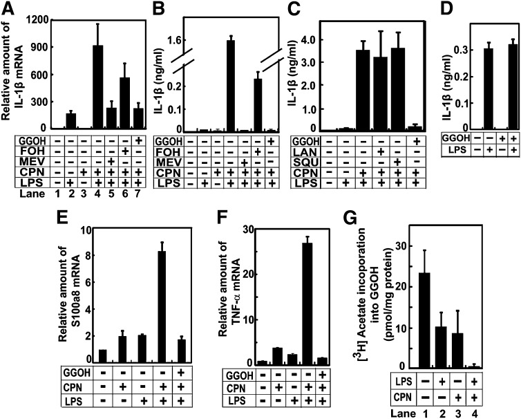
Fig. 4.
GGOH is required to maintain endotoxin tolerance. A–C, E, F: On day 0, peritoneal macrophages were seeded at 5 × 10⁶ cells per 60 mm dish. On day 1, cells were switched to medium containing (+) or not containing (−) 200 ng/ml LPSs, 25 μM compactin (CPN), 250 μM mevalonate (MEV), 10 μM FOH, GGOH, squalene (SQU), and lanosterol (LAN) as indicated. On day 2, 24 h after the first treatment, cells were switched to fresh medium that was used in the first treatment. On day 3, 24 h after the second treatment, cells were harvested for quantification of indicated mRNA as described in Fig. 3A (A, E, F). The amount of IL-1β protein secreted into medium was determined as described in Fig. 3B (B, C). D: On day 0, peritoneal macrophages were seeded at 5 × 10⁶ cells per 60 mm dish. On day 1, cells were treated with 10 μM GGOH as indicated for 24 h. On day 2, cells were switched to the same medium with or without the addition of 200 ng/ml LPSs as indicated. On day 3, 24 h later, the amount of IL-1β protein secreted into medium from macrophages exposed to LPSs for the first time was determined as described in Fig. 3B. G: On day 0, peritoneal macrophages were seeded at 5 × 10⁶ cells per 60 mm dish. On day 1, cells were switched to medium containing 200 ng/ml LPSs and 25 μM compactin as indicated. Eight hours after the treatment, cells were switched to the identical medium containing 10 μCi [3H]acetate and 0.5 mM unlabeled acetate. The amount of the radioactivity incorporated into GGOH was determined as described in Experimental Procedures. A–G: Results are presented as mean ± SE from three independent experiments.

