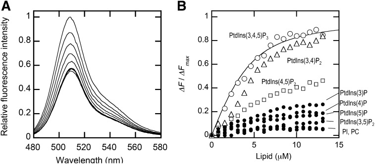
Fig. 3.
Fluorescence quenching of the C-terminal EGFP-tagged PDK1 PH domain (PDK1-PH-EGFP) by dabsyl-PE-containing vesicles. A: Fluorescence emission spectra of PDK1-PH-EGFP (100 nM) in the presence of increasing concentrations of POPC/POPS/dabsyl-PE/PtdIns(3,4,5)P3 (72:20:5:3) vesicles were measured in the cuvette-based assay. The total lipid concentrations used were 0, 1.6, 3.9, 5.4, 6.9, 8.5, 9.2, 10.0, and 11.6 μM from top to bottom. Fluorescence emission intensity was normalized against the EGFP intensity value at 509 nm without lipid vesicles. B: Binding isotherms of PDK1-PH-EGFP with vesicles containing various PtdInsPs, i.e., POPC/POPS/dabsyl-PE/PtdInsP (72:20:5:3) vesicles. PtdInsP includes PtdIns(3,4,5)P3, PtdIns(3,4)P2, PtdIns(4,5)P2, PtdIns(3,5)P2, PtdIns(3)P, PtdIns(4)P, PtdIns(5)P, and PtdIns. POPC/dabsyl-PE (95:5) vesicles were also tested as a negative control. The fluorescence decrease upon lipid addition was background corrected by the fluorescence decrease upon addition of the same volume of the buffer solution. ΔF/ΔFmax was calculated as fluorescence intensity decrease at a given lipid concentration divided by the maximal fluorescence decrease. The binding isotherm for POPC/POPS/dabsyl-PE/PtdIns(3,4,5)P3 (72:20:5:3) vesicles was fit by nonlinear least-squares analysis using equation 1. Values of n (40 ± 10) and Kd (35 ± 10 nM) are summarized in Table 1.

