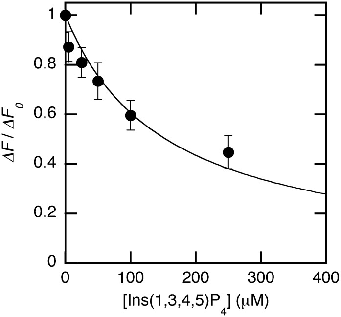
Fig. 6.
Inhibition assay for membrane binding of PDK1-PH-EGFP. Each row of a 96-well plate contained a fixed concentration of Ins(1,3,4,5)P4, a fixed concentration of PDK1-PH-EGFP (100 nM), and the increasing concentration of POPC/POPS/dabsyl-PE/PtdIns(3,4,5)P3 (72:20:5:3) vesicles (0 to 25 μM). The maximal fluorescence decrease for each concentration of Ins(1,3,4,5)P4 (ΔF) was then determined and converted into ΔF/ΔFo. ΔFo is the maximal fluorescence decrease in the absence of Ins(1,3,4,5)P4. A control row contained the buffer solution. IC50 values were determined by the nonlinear least-squares analysis using equation 2. Each point shows the mean ± SD value from triplicate measurements.

