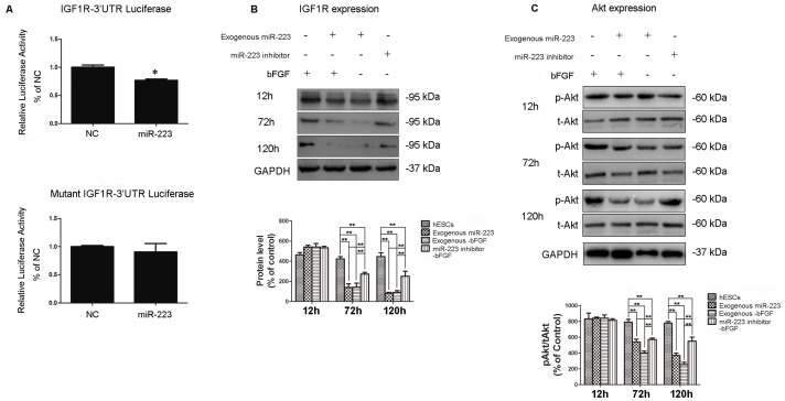
Figure 3. MiR-223 regulates hESCs differentiation likely via IGF-1R/Akt.
(A) hESCs were infected with lentiviruses expressing miR-223 and wild type IGF-1R-3′UTR or a mutant IGF-1R-3′UTR construct. After 48 h, luciferase (Luc) activity was assayed. Data are presented as the mean ± SEM of the percentage of luciferase activity detected in control cells (*p<0.05; n = 4). Western blotting was performed to detect levels of IGF-1R (B) and phosphorylated Akt (p-Akt) and total Akt (t-Akt) (C) 12, 72, and 120 h after withdrawal of bFGF for the indicated groups. NC indicates the mock-vehicle group and GAPDH was used as a loading control. Protein bands were semi-quantified using imaging analysis software. Data are expressed as the mean ± SD and are representative of the three independent experiments that were performed. *p<0.05, **p<0.01, n = 3.

