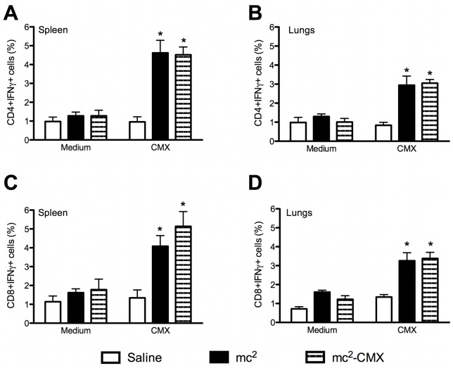
Figure 3. Induction of specific cellular responses to ex vivo stimulation with CMX.
Lung and spleen single-cell suspensions from vaccinated and control mice were restimulated with CMX or medium, and the CD4+ and CD8+ IFN-γ-positive T cells were analyzed by flow cytometry. Lymphocytes were selected by their size and granularity and the CD4+ cells were gated and analyzed for IFN expression. A and B show CD4+ IFN-γ-positive cells from the spleen and lungs, respectively. C and D show CD8+ IFN-γ-positive cells (n = 4; *p<0.05) from the spleen and lungs, respectively. The results are representative of two independent experiments.

