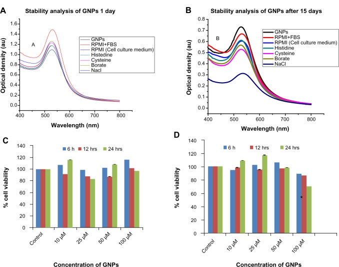
Figure 4.
Stability and cytotoxicity of GNPs (top panel). UV-vis spectra of the GNPs in various buffers at various time periods: (A) 1 day; (B) 15 days. Cytotoxicity measurements of GNPs in different cell lines at various concentrations (bottom panel): (C) MIO-M1 (Müller glial, noncancerous); (D) MDA-MB 453 (breast cancer). The cell viability (% of treated cells with respect to untreated cells) of different cell lines treated with different concentrations: 10, 25, 50, and 100 μM of GNPs at various time periods.
Notes: *Significant difference with respect to control at P< 0.05; error bars represent the standard error of mean.
Abbreviations: GNPs, gold nanoparticles; RPMI+FBS, Roswell Park Memorial Institute medium plus fetal bovine serum; RPMI, Roswell Park Memorial Institute medium; Nacl, sodium chloride; UV-vis, ultraviolet-visible; au, arbitrary unit.

