Figure 2.

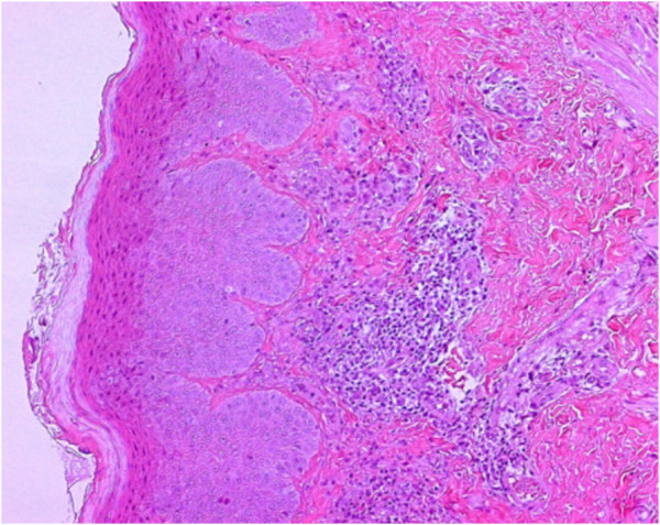
Histological image of the epidermal layer with a mild lymphohistiocytic and granulocytic infiltrate, predominantly eosinophilic.

Histological image of the epidermal layer with a mild lymphohistiocytic and granulocytic infiltrate, predominantly eosinophilic.