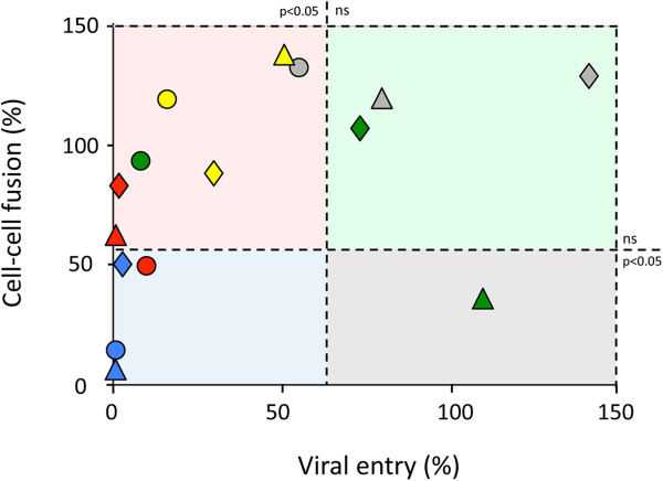
Figure 5.
Correlation between cell-cell fusion and viral entry for envelopes carrying an exogenous V1, V2, or V1V2 region. Values are the average of 3 to 5 independent experiments for viral entry assays or the average of 3 independent experiments for cell-cell fusion assays. The graph is divided into four sections according to whether the proteins located in one quarter displayed or did not display a level of functionality significantly lower than that of the corresponding wild-type protein in viral entry or in cell-cell fusion assays. The threshold of significance is set at p < 0.05, as indicated in the figure (ns = not significant). The bars indicating the threshold of significance are drawn in the median position between the position of the last sample that has a significant decrease in functionality and the first one that does not display a significant difference (gray circle and green diamonds for cell entry; blue diamonds and red triangle for cell-cell fusion). V1V2 chimeras, diamonds; V1 chimeras, circles; V2 chimeras, triangles. Chimeras: ABA, gray, ACA, yellow; AGA, green; BCB, red; BGB, blue.

