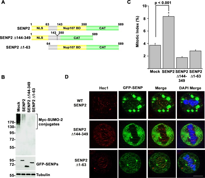
FIGURE 5:
N-terminal targeting elements in SENP2 specify mitotic arrest phenotypes. (A) Schematic diagram of SENP2 and targeting domain mutants. BD, binding domain; CAT, catalytic domain; NLS, nuclear localization signal. (B) HeLa cells were cotransfected with constructs coding for Myc-tagged SUMO-2 and wild type GFP-SENP2, the indicated GFP-tagged SENP2 mutants, or empty vector as control (Mock). Cell lysates were analyzed by immunoblot analysis with antibodies specific for Myc, GFP, or tubulin. (C) HeLa cells were transfected with constructs coding for wild-type GFP-SENP2, the indicated GFP-tagged SENP2 mutants, or empty vector (Mock). The fraction of transfected cells in mitosis was determined by fluorescence microscopy 48 h after transfection. (D) HeLa cells were transfected with constructs coding for wild-type GFP-SENP2 or the indicated GFP-tagged SENP2 mutants. Cells were permeabilized, fixed, and stained with Hec1-specific antibodies and analyzed by immunofluorescence microscopy. DNA was labeled with DAPI. Bar, 10 μm. Error bars represent SDs from three independent experiments.

