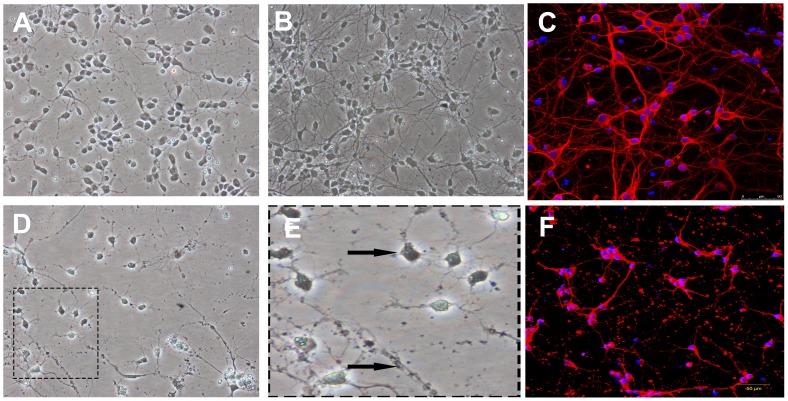
Figure 3. Morphology of primary rat cortical neurons and OGD injury.
(A) 200× First day in vitro, neurons were small and adhered to bottom of the flask to grow, with a round body and small neurite. (B) 200× Fifth day in vitro, these neurites of neurons formed an extensive network. (C) Immunofluorescence shows cell body and neurite were labeled by anti-class III β-Tubulin antibody (red), while nuclei were stained with Hoechst33342 (blue). Scale bar = 50 µm. (D) 200× After 24 h, neurite of neurons following by 90 min of OGD injury were degraded and disappeared, with partial dead cell. (arrowhead shown in Fig. E) (magnification). (F) Immunofluorescence shows cell body and neurite after OGD injury. Scale bar = 50 µm.

