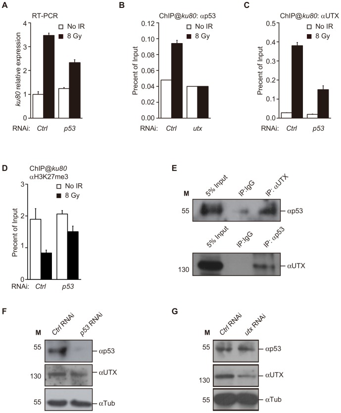
Figure 3. p53 and UTX are recruited in an interdependent manner to the ku80 promoter region.
(A) ku80 expression following IR exposure in Kc cells subjected to RNAi treatment, as indicated. (B, C) ChIP analysis of the physical occupancy of p53 and UTX at the ku80 promoter region. Note that knockdown of utx eliminates the increase in p53 binding, and knockdown of p53 reduces the binding of UTX to the ku80 promoter. (D) ChIP assay for H3K27me3 at the ku80 promoter in Kc cells treated with control or utx RNAi after IR. (E) Coimmunoprecipitation was performed using anti-p53 and anti-UTX antibodies and whole cell extracts of Kc cells. The immunoprecipitates were subjected to Western blot analysis with the indicated antibodies. (F, G) Western blot analysis to confirm the knockdown efficiency of p53 RNAi. β-Tubulin (β-Tub) levels were used as a loading control.

