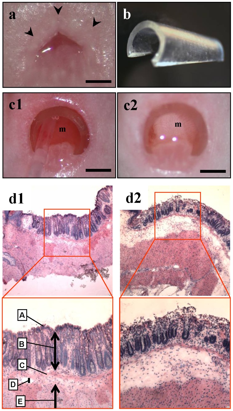
Figure 1. Disruption of the mucosal barrier of the rectum.

(a) Normal appearance of mouse anus. Scale bar, 1 mm. (b) Retractor made from a drip infusion tube. (c) The anorectal lumen was dilated by inserting the retractor into the anorectum and instilled with an acetic acid solution. (c1) Before acetic acid preparation. (c2) The color of the rectal mucosa changed from reddish pink to whitish after treatment with acetic acid solution. m = rectal mucosa. Scale bar, 1 mm. (d) Histological examination just after acetic acid treatment showed that the epithelial cell layer of the rectal mucosa was traumatized. (d1) H&E section of normal anorectum. A = surface epithelium; B = mucosa; C = muscularis mucosae; D = submucosa; E = muscularis externa. (d2) After acetic acid treatment. Note that only the upper part of the mucosa is disrupted. Top, × 40 magnification; bottom, × 100 magnification.
