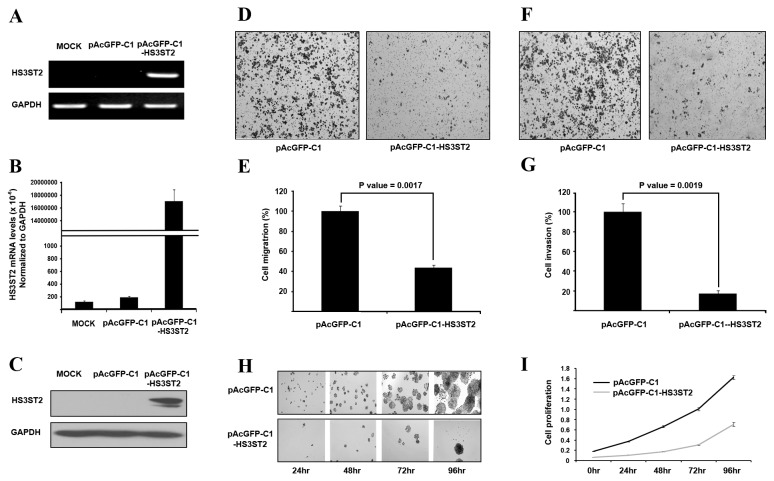
Figure 3. Induced expression of HS3ST2 inhibited cell migration, invasiveness, and proliferation.
(A-C) pAcGFP-C1-HS3ST2 was transfected into H460 lung cancer cells. Specific overexpression of HS3ST2 compared to the empty vector was confirmed by RT-PCR (A), qRT-PCR (B), and western blotting (C). (D-G) H460 cells were seeded in a Boyden chamber with an empty vector or with one μg pAcGFP-C1-HS3ST2 in transwell migration (D & E) and invasion assays (F & G) as described in the Materials and Methods. Stained migrating cells and invading cells are shown in (D) and (F), respectively; migrating cells were measured by absorbance at 564 nm; invading cells were counted under a microscope. The results are presented relative to the in vitro migration or invasion of uninduced H460 cells (100%). (H & I) H460 cells transfected with an empty vector or pAcGFP-C1-HS3ST2 were cultured in a 96-well dish to analyze the effect of HS3ST2 on cell growth. Cell proliferation was measured by MTT assay and absorbance was measured at 570 nm. Data are presented as the mean ± standard error (SE) of triplicate experiments.

