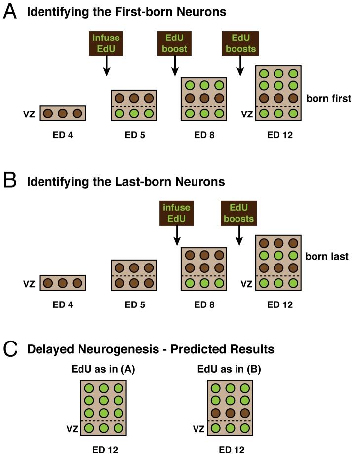
Figure 2. Schematic of the experimental design and predictions.
On either ED5 (A) or ED8 (B), control and FGF2-treated embryos were infused with EdU followed by regular booster shots sufficient to saturate the system. Birds were then sacrificed on ED12 for processing. EdU is taken up by all proliferating cells as they pass through S-phase (shown in green), whereas all cells born prior to infusion are EdU-unlabeled (shown in brown). In the ED5 condition (A), the early born neurons in the deep layers of control tecta have already been born at the time of EdU infusion and so do not take up EdU (brown). In contrast, if FGF2 delays neurogenesis, then all cells should take up the EdU (green) in the treated embryos (C), because no neurons would have been born at the time of EdU infusion. In the ED8 condition (B), only late born neurons in the middle layers of control tecta have yet to be born, and so take up the EdU (green). If FGF2 delays tectal neurogenesis, then one would expect the neurons in the most superficial layers also to incorporated the EdU, because they, too, would not yet have been born before the EdU infusion (C). The ventricular zone (VZ) contains proliferating cells and is, therefore, EdU-positive after the EdU infusions have begun.

