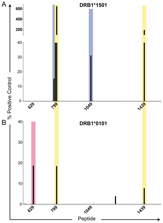
Figure 3. α1(V) peptide binding to HLA-DR1 and HLA-DR15.
ProImmune REVEAL MHC-peptide binding assay screen of 5-amino acid overlapping peptide library (101 total peptides) of the α1(V) helical domain to HLA-DR15 (A), and HLA-DR1 (B). Each MHC:peptide binding score is based on known binding of a positive control peptide and a weaker binding positive control peptide. MHC:peptide binding scores above 12% are considered positive and have potential for immunological activity. Lack of bar for a peptide represents a score of 0%. Peptide number corresponds to the location in the α1(V) AA sequence. Blue shaded areas indicate epitope regions where only HLA-DR15 bound, red shaded areas where only HLA-DR1 bound, and yellow shaded areas indicate both HLA-DR1 and –DR15 bound within that helical domain.

