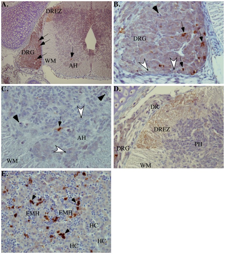
Figure 3. Micrographs of immunohistochemical analyses of fetal tissues with 1277 neoepitope antisera detecting the active p20 subunit of Casp6.
A. At low power (original magnification, 100x), immunopositive cells (arrows) in the dorsal root ganglion (DRG), in the anterior horn (AH), and in the dorsal root entry zone (DREZ) of the spinal cord white matter (WM). B. High power view of dorsal root ganglion (DRG: original magnification, 400×) indicating mitotic activity (solid arrowhead), features of early neuronal differentiation (open arrowheads) and several apoptotic immunopositive cells (arrows). C. High power view of anterior horn (AH: original magnification, 400×) indicating dividing cells (solid arrow heads), early maturation of cells (open arrowheads), and an immunopositive apoptotic precursor cell (arrow). WM; white matter). D. High power view of dorsal spinal cord (original magnification, 400×). Immunopositive finely granular (synaptic) pattern in the dorsal root entry zone (DREZ) of the spinal cord white matter (WM). DRG, dorsal root ganglion; DR, dorsal root; PH, posterior horn). E. Extensive extramedullary hematopoiesis (EMH) in the sinusoids that separate the cords of hepatocytes (HC) in the liver (original magnification 400×). In the hematopoietic islands, there are many immunopositive apoptotic cells (arrows).

