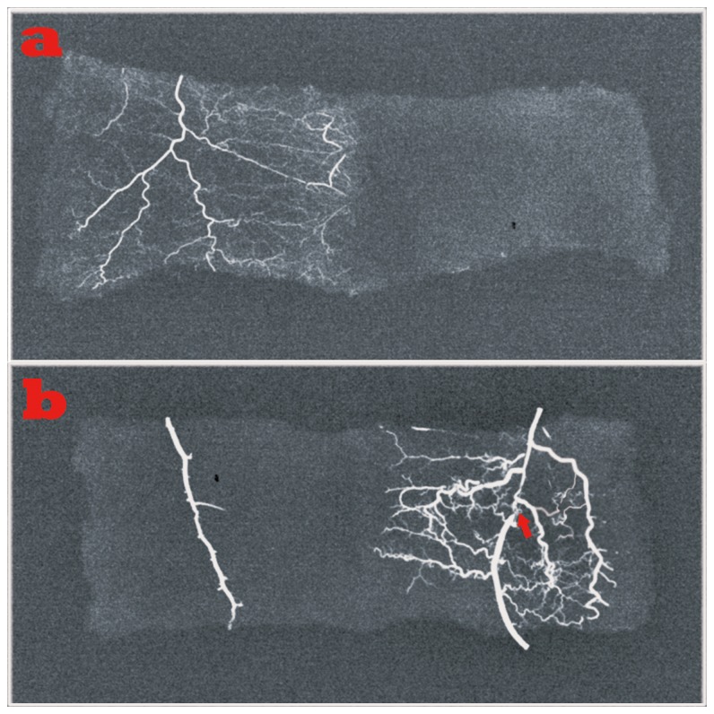
Figure 5. Microvasculature examination of functional vessels.
a. Left, dense vessels were distributed regularly around the flaps of the arterial perfusion group; right, no vessels were visible in flaps of the composite skin-grafting group.
b. Left, only a trunk vessel without small vessels was noted in the AVF group; right, plenty of small vessels as seen in the arterial perfusion group was visible in HR group ( the red arrow indicates the site of ligation).

