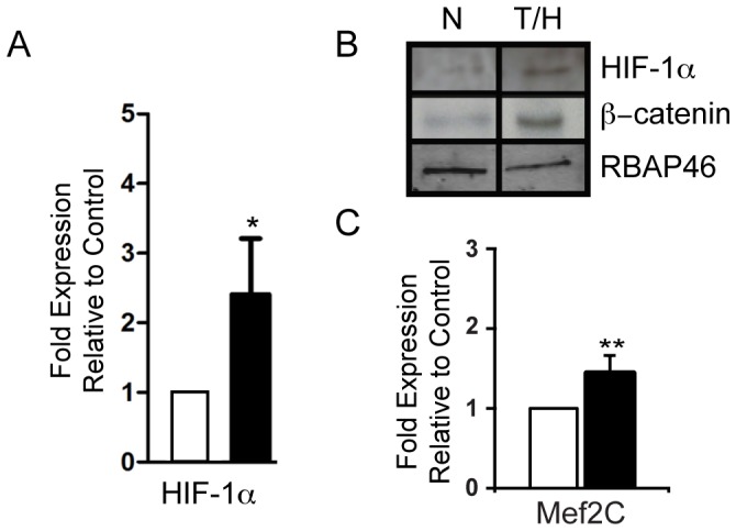
Figure 5. Effects of hypoxia on HIF-1alpha and beta-catenin expression.

A. Quantitative PCR showing significant increase of HIF-1α expression following 24 hours hypoxia alone compared to normoxic control. B. Representative Western blots of nuclear extracts from EBs, following 24 h or normoxia or hypoxia. Protein loading was examined by Western blotting against the nuclear protein RBAP46. C. Quantitative PCR showing significant increase of Mef2c expression following 24 hours hypoxia alone compared to normoxic control. All qPCR experiments (n = 4) were performed in 2 independent experiments.
