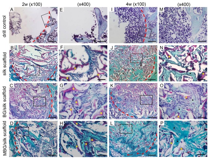
Figure 8. Saffranin O staining of bone formation (black star) and scaffolds remnant (yellow star) within defects in the drill control, silk, BG/silk and MBG/silk groups at 2 and 4 weeks.
Traces of cartilage matrix (red arrow head) can be observed in MBG/silk groups. Lower magnification (x100; A-C, I-L; bar=200 µm); higher magnification (x400; E-H, M-P; Bar=50µm). The red dotted line indicated defect margin.

