
Figure 5. The OVAL locus is focally amplified in serous endometrial tumors.
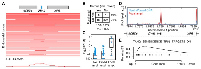
A, Low frequency focal amplification of the OVAL locus in endometrial cancer, but not 16 other TCGA cancers (see Figure S6 in File S1). B, 56% of focally amplified cases were of the serous subtype, compared to 21% overall (P = 0.025, Fisher’s exact test). C, OVAL RNA was strongly induced in a subset of tumors, and this coincided with focal amplification of the AXI region. ND, not detected. D, Average RNA-seq read density in the AXI region for tumors with marked focal amplification (n = 4) compared to remaining tumors (normalized read counts per 1000 nt segment). E, Similar to ovarian cancer, GSEA analysis revealed induction of P53 targets in OVAL amplified tumors.