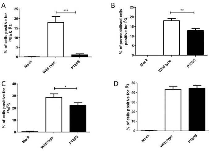
Figure 2. Expression of normal and mutated αIIbβ3 and αvβ3 in CHO cells.
CHO cells were transfected with either wild type or mutated β3 P163S expression plasmids alone (C, D) or in the presence of wild type αIIb expression plasmid (A, B), or mock transfected with empty vector as a negative control. Forty-eight hours after transfection, the percentage of cells expressing both αIIb and β3 (A), αvβ3 (C) and β3 (D) were determined by flow cytometry. Intracellular expression of β3 was assessed after permeabilization of the cells (B). Data represent the mean and standard deviation of three independent experiments. ***p<0.001, **p<0.01, *p<0.05 as calculated by unpaired t-tests.

