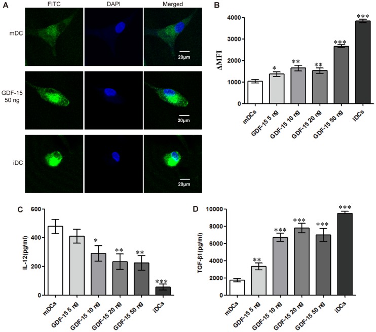
Figure 5. GDF-15 affects phagocytosis and cytokine secretion by DCs.
(A) Confocal microscopy. CD14+ cells were cultured in a 15 mm confocal dish and induced to form DCs under different culture conditions. After incubation with FITC-dextran, fixation and staining with DAPI, the cells were observed by confocal microscopy. Original magnification ×400. The scale bars are equal to 20 μm. (B) Flow cytometry. The results are shown as the ΔMFI. For each sample, the background (MFI of the fluorescence of the cells pulsed at 4°C) was subtracted from the MFI of the cells incubated at 37°C. (C, D) Levels of cytokine secretion in DCs. The culture medium was collected when the cells were harvested for analysis. After centrifugation, IL-12 (C) and TGF-β1 (D) levels in the supernatants were detected using an ELISA kit according to the manufacturer's instructions. The OD450nm was recorded using a spectrophotometer. Mean ± SD, n = 3. * P<0.05, ** P<0.01 and *** P<0.001 compared with the mDCs. The experiments were conducted in triplicate.

