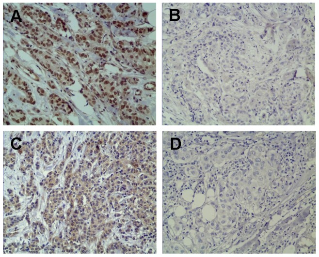
Figure 1. Representative immunohistochemistry images.

MDM2 expression was demonstrated by brown-staining in the cell membrane or cytoplasm (A), while MMP-9 expression was shown as brown-staining in the nucleus or cytoplasm (C). The negative staining patterns are presented to enable a comparison (B, MDM2 negative, and D, MMP-9 negative).
