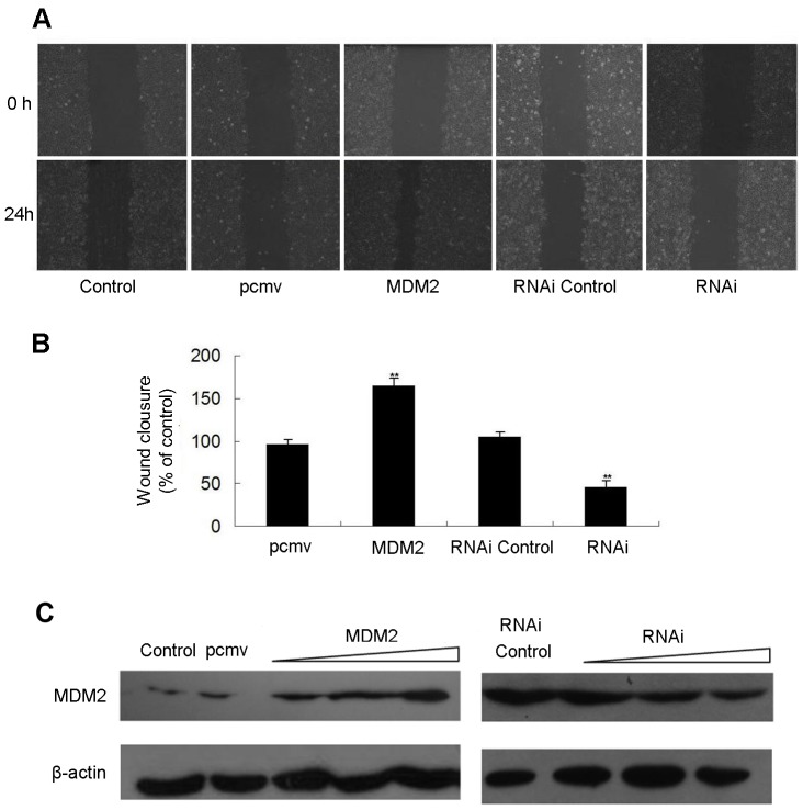
Figure 5. MDM2 promotes the migration of MCF-7 cells.
MCF-7 cells were transfected with pcmv-MDM2 expression plasmids and pcmv vectors or siRNAs against MDM2 and non-specific siRNA (controls); after 24 h, the cells were scraped with a sterile pipette tip to create a wound. (A) Wound closure was observed by phase-contrast microscopy and photographed at 0 and 24 h. (B) The wound area was measured by the Adobe Photoshop software. Wound closure was quantified as the mean ± standard deviation of three independent experiments. The control wound closure was set at 100%, and the MDM2 treatments are represented as the percent of the control. (C) The levels of MDM2 protein were detected using Western blot analysis in MCF-7 cells that over- or under-expressed MDM2 (plasmid or siRNA transfection) and control cells. β-actin levels served as internal control.

