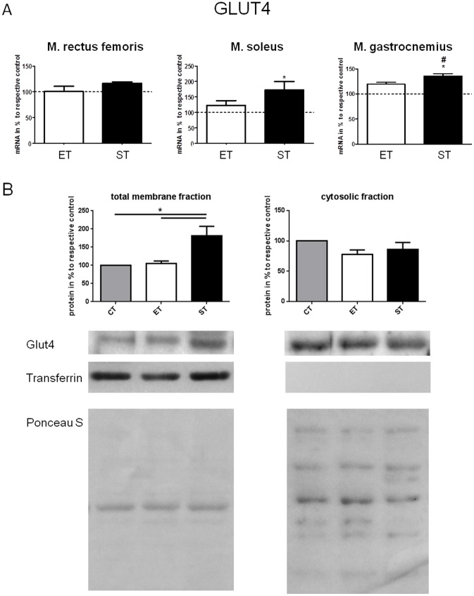
Figure 6. A. Relative expression of mRNA of GLUT4 in m. rectus femoris, m. soleus, and m. gastrocnemius of ET and ST group relative to respective control.
Values of CT group were set to 100%. */# indicates significant differences to control and ET respectively (p<0.05). B. Expression of GLUT4 proteins in total membrane and cytosolic fraction of m. rectus femoris in mice of the CT, ET and ST group. Values of CT group were set to 100%. Gels were spliced to present the results from the strength training (ST) in the consecutive sequence according to figures. * shows significant differences as indicated (p<0.05). Ponceau S staining demonstrates equal loading of proteins. Data are presented as mean ± SEM.

