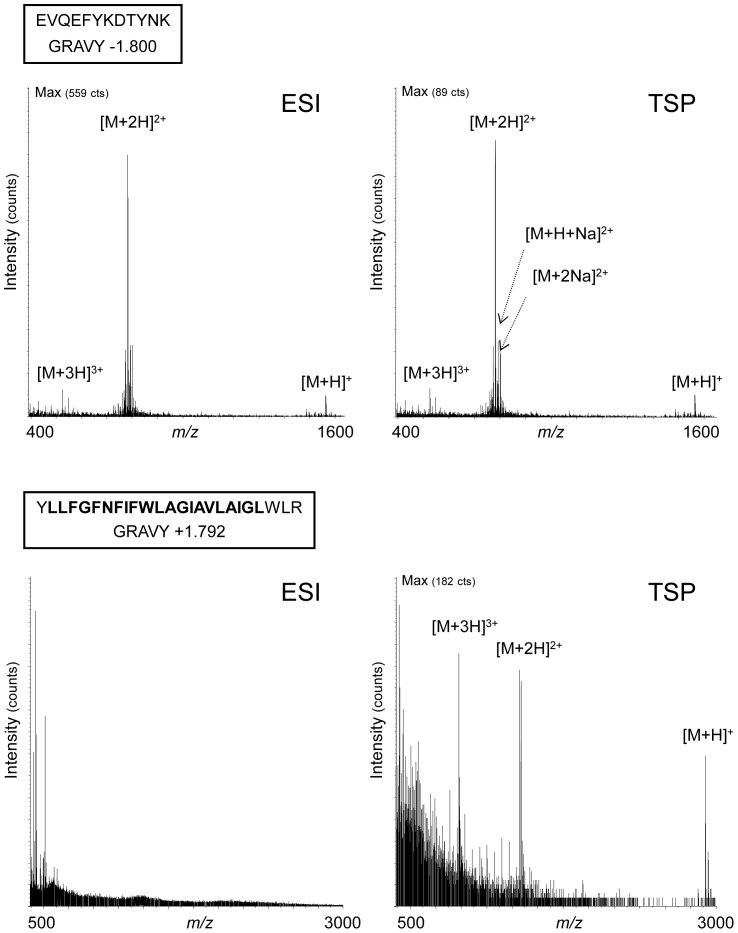
Figure 1. Ionization of two extreme hydrophilic and hydrophobic peptides in ESI or TSP.
The hydrophilic peptide located in the second extracellular loop of the protein CD9 (aa 120–131) (upper panel) and the highly hydrophobic peptide corresponding to the first transmembrane domain (aa 12–36) (lower panel) were investigated by mass spectrometry using electrospray ionization (ESI) or thermospray (TSP) as ionization modes. The sequence corresponding to the transmembrane domain is in bold.

