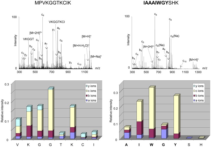
Figure 4. In-source fragmentation of peptides under APPI.
Two peptides originating from the protein CD9 corresponding to a hydrophilic peptide (aa 1–11, Gravy −0.155) (left) and a hydrophobic peptide (aa 104–114, Gravy +0.355) (right) were investigated by APPI under dopant assisted conditions using toluene and 9 eV photons. The sequence embedded in the membrane is represented in bold. Along with precursor ions, abundant and intense fragment ions were detected in the mass spectra corresponding to in-source fragmentation of studied peptides. The intensities of each fragment species a-, b-, c- and y-ions were plotted in relation to the peptide sequence.

