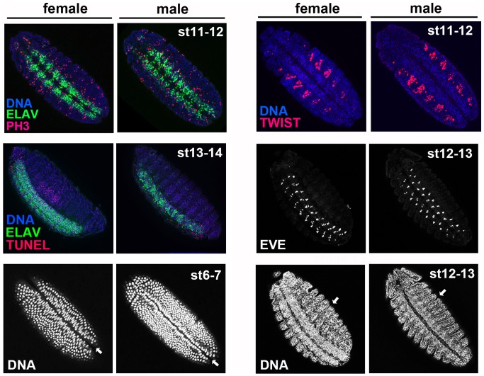
Figure 3. Spiroplasma does not cause abnormal levels of cell proliferation, cell death, or abnormal defects in non-neural tissues or other processes during the onset of neural defects.
All embryos shown are MSRO-infected. Cell proliferation is depicted by anti-phospho histone H3 (PH3) (red in top left panels); broken DNA, an indicator of cell death, is shown by TUNEL (red in middle left panels); The ventral midline invagination can be seen with DNA staining (indicated by white arrows in bottom left panels); The developing mesoderm is highlighted by anti-Twist (red in top right panels); A subset of motoneurons located in odd body segments is marked by anti-even-skipped (white in middle right panels); external body segments are seen through DNA staining (indicated by white arrows in bottom right panels).

