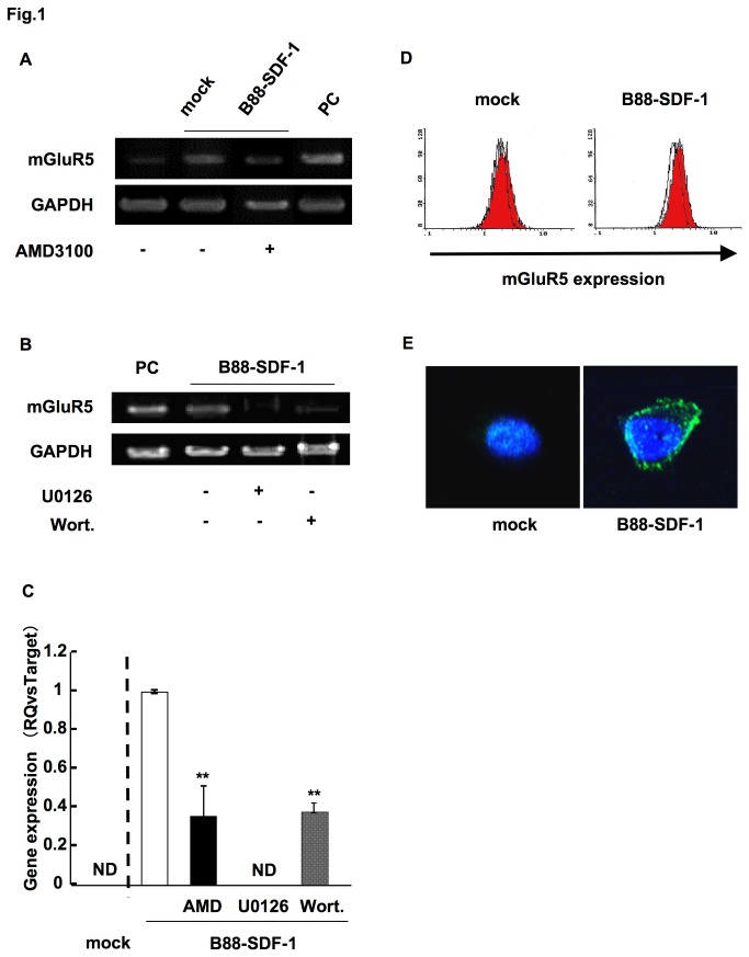
Figure 1. The upregulation of mGluR5 in B88-SDF-1 cells.
(A) Expression of mGluR5 mRNA was confirmed in B88-mock and B88-SDF-1 cells in both the presence and absence of AMD3100 (1 µg/ml). Human placenta was used as a positive control (PC). (B) Cells were treated with U0126 (10 nM) or wortmannin (50 nM) for 48 h and mRNA expression of mGluR5 was analyzed by RT-PCR. (C) Expression of mGluR5 mRNA was confirmed by the real-time PCR. **; p < 0.01 when compared to untreated B88-SDF-1 cells by one-way ANOVA. ND; not detectable. (D) Protein expression of mGluR5 was evaluated in B88-mock and B88-SDF-1 cells using flow cytometry. Logarithmically growing cells were incubated with or without anti-mGluR5 mAb and stained with PE-labeled goat anti-mouse IgG. White and red zones indicate cells stained with the isotype control and the anti-mGluR5 mAb, respectively. (E) Protein expression of mGluR5 was detected by immunocytochemistry. The nucleus was stained with DAPI (blue).

