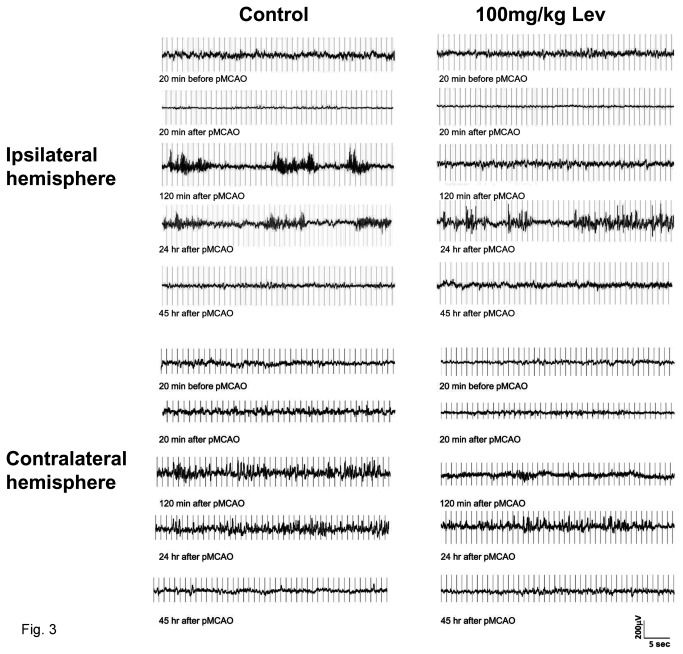
Figure 3. Effect of brain ischemia and of Levetiracetam on EEG activity.
The figure reports EEG recording samples of about 40 sec in duration obtained in a representative control (on the left) and a Lev-treated rat (on the right) before and after pMCAO. For each animal, traces obtained immediately before, and 20 min, 120 min, 24 and 45 hours after pMCAO are reported. The traces shown in the panels on the top of the figure were recorded ipsilaterally to MCAO whereas those in the bottom panels are from the contralateral side. Note that EEG activity was markedly suppressed after pMCAO in the ipsi- but not in the contralateral brain hemisphere. Note also that a significant EEG activity reappeared, in the form of NCSs, 120 min after pMCAO in the control rat and only 24 hours after vessel occlusion in the Lev-treated rat. In the traces from the contralateral hemisphere reported in the bottom panels, IRDAs can be easily identified as brief bursts occurring in isolation. Note that these events are already large and well defined in traces obtained 120 min after pMCAO in control but not in Lev-treated rats in which they become clearly evident only after 24 hours.

