
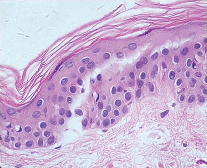
Figure 6.
Histological features of vitiligo that on the edge of the skin lesions by RCM can be seen highly refractile inflammatory cells showed (H and E, ×400): Hypopigmentation, no lymphocytic infiltration
Official websites use .gov
A
.gov website belongs to an official
government organization in the United States.
Secure .gov websites use HTTPS
A lock (
) or https:// means you've safely
connected to the .gov website. Share sensitive
information only on official, secure websites.
Histological features of vitiligo that on the edge of the skin lesions by RCM can be seen highly refractile inflammatory cells showed (H and E, ×400): Hypopigmentation, no lymphocytic infiltration