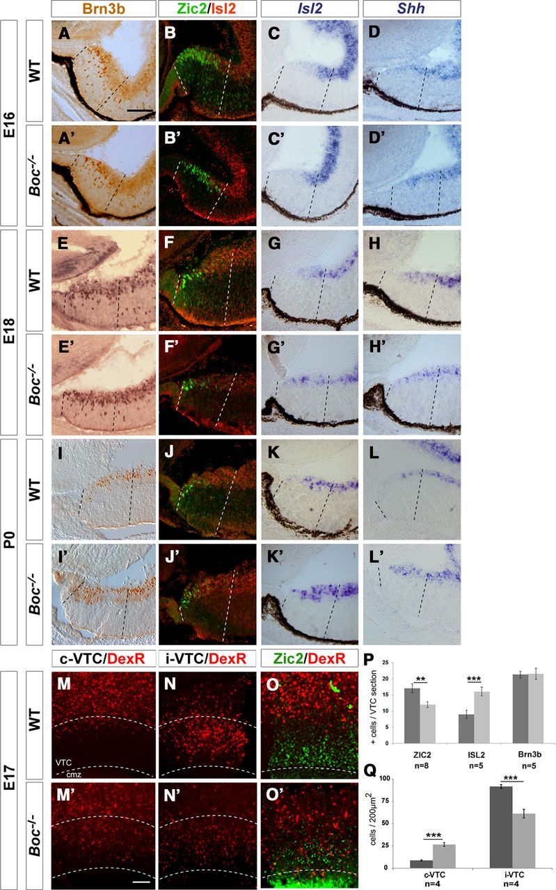
Figure 5.

A–L′, Boc is required to restrain Islet2/Shh expression within the VTC. Frontal cryostat sections from E16 (A–D′), E18 (E–H′), and P0 (I–L′) WT and Boc−/− central retinas immunostained for Brn3b, double-stained for Zic2 (green) and Islet2 (red), or hybridized with specific probes for Isl2 and Shh, as indicated in the panels. The increase in Islet2+ cells parallels the decrease in Zic2+ cells in Boc−/− tissue. There is a correspondent increase in Islet2 and Shh expression in the mutants starting from E18. Dashed lines delimit the VTC. M–O′, Low-power views of flat-mounted preparations of the ipsilateral and contralateral retinas from E17 embryos subjected to unilateral retrograde labeling from the optic tract, double-labeled with antibodies against Zic2 (O,O′). In WT, the majority of backfilled neurons localized to the ipsilateral VTC (the VTC area is marked with dotted lines), whereas only few RGCs were found in the contralateral VTC. In Boc−/− VTC, this proportion is shifted and there are numerous Zic2+ backfilled cells in the cVTC (O′). P, Q, The histograms represent the amount of Zic2+, Islet2+, or Brn3b+ cells in P0 (P) and the amount of backfilled cells present in the ipsilateral and contralateral retinas of E17 (Q), and WT (dark gray bars) and Boc−/− (light gray bars) VTC. **p < 0.01 (Student's unpaired t test). ***p < 0.001 (Student's unpaired t test). cmz, Ciliary marginal zone. Scale bar, 100 μm.
